![]()
![]()


INTRODUCTION TO THE HUMAN BODY
Definitions
Anatomy deals with the structure and the relationships among the various
structures of the body. Physiology deals with how the body works.
Rise of the Scientific
Method
In an effort to support
natural phenomena other than a mixture of belief, superstition, and argument a
method had to be developed that was not based on prejudice. Two types of
reasoning are applied; an inductive approach whereby the
scientist accumulates data and then formulates a hypothesis to account for those
facts; and a deductive approach whereby the scientist constructs
a hypothesis, tests its validity outlining particular events that are predicted
by the hypothesis, and then performs experiments to test for those events. In
time hypothesis gives rise to a theory (a collection of statements or concepts
that explains a natural phenomenon. At some point in time a theory becomes a
law. Most physiological knowledge was obtained by the hypothetico-deductive
method. Usually a hypothesis is in an "if-then" statement. A researcher will
state, "If my hypothesis on ____ is correct, and I record my observations during
the ____ experiment, then I should observe _____ results." The researcher will
design her experiment with enough of a sample size to generate meaningful
results, include a control group along with the test group to see if the
experiment produced any differences. Results will be analyzed statistically to
determine differences.
Human Structure
The Levels of structural organization from smallest to largest are Chemical
(atoms and molecules), Cellular (basic units of the organism), Tissue (groups of
similar cells that function together), Organ (structures that are composed of
two or more tissues, have specific functions, and usually have a recognizable
shape), and System (groups of related organs that have a common function).
|
Characteristics of Life |
|
| Organization | Living things exhibit a higher level of organization than nonliving things and spend a good deal of energy maintaining that organization. |
| Cellular composition | Always compartmentalized into one or more living cells |
| Metabolism & Excretion | Living things take in molecules, change those molecules to aid them in living and excrete waste materials from the chemical reactions that occur. |
| Responsiveness & movement | This refers the to ability to sense and react to stimuli throughout all levels of the body. Living things have the ability to propel themselves from place to place. |
| Homeostasis | Maintenance of a stable internal environment no matter what the external environment is doing. |
| Development | Compose of differentiation of cells into specialized organs and growth in size. |
| Reproduction | Being able to produce a copy of oneself. |
| Evolution | Exhibit genetic change from generation to generation. |
When one thinks about it, none of the
chemical constituents of our bodies is alive. Put them together and they still
are not alive. What then is life? The Characteristics of Life that determine whether or not an
organism is alive are listed in the table above.
Life is not a single property, but a collection of properties that distinguish
it from nonliving things. Organisms do not have to these things at the same time, but should have those
capabilities at sometime during their life cycle.
The Requirements of organisms to survive are water, food, oxygen, heat, and
pressure.
Homeostasis
Homeostasis is a condition in which the body’s internal environment, which
is very dynamic, remains
within certain physiological limits. Homeostasis contains the optimum
concentration of gases, nutrients, ions, and water. Homeostasis has an optimum
temperature. Homeostasis has an optimum volume for the health of the cells.
Stress is a factor the affects homeostasis. Throughout this course, we will
continue to come back to the theme of homeostasis.
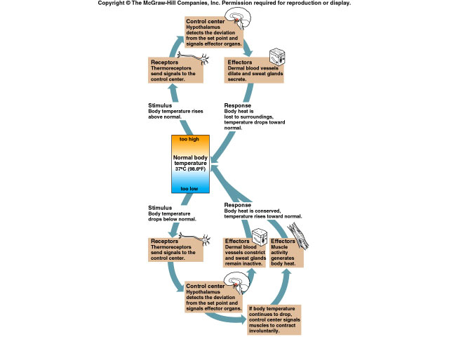
Regulation of homeostasis is done by the nervous and endocrine systems either working together or separately. Homeostasis depends on a negative feedback system. A negative feedback system reverses the original stimulus whereas positive feedback system enhances the original stimulus.
-
The nervous system, via receptors, monitors changes and the status of the body and send inputs to the control center.
-
A control center determines the point at which a controlled condition should be maintained.
-
If necessary, effectors receive information from the control center and produce a response. The endocrine system releases hormones to maintain homeostasis. The muscular system produces a movement. One thing is certain; disruption of homeostasis can lead to disease and death.
ORIENTATION OF THE BODY
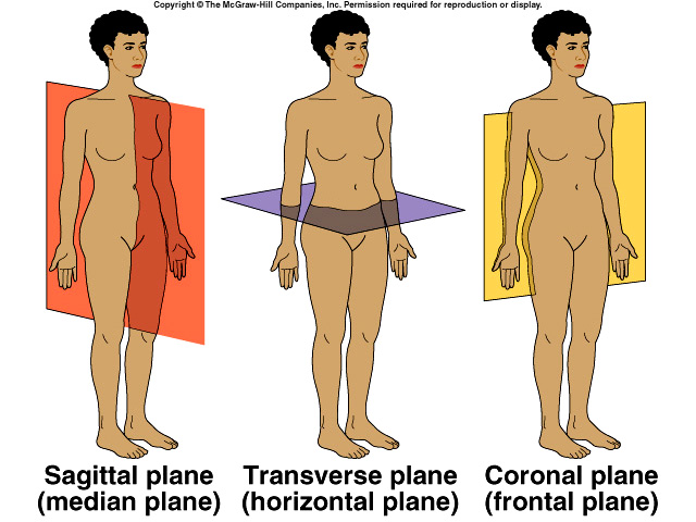
ANATOMICAL POSITION
The anatomical position is a stance in which a person stands erect with feet
flat on the floor, arms at their sides, and palms, face, and eyes facing
forward.
PLANES OF THE BODY
Midsagittal or median: vertical plane that
divides the body or organ in two.
Parasagittal: vertical plane that divides the body or organ into two unequal
parts.
Frontal: divides body or organ into anterior and posterior portion.
Horizontal (transverse) divides body or organ into equal superior and inferior
portions.
Oblique: passes through body or organ at an angle.
DIRECTIONAL TERMS: indicate relationships of one part of the body to another
Superior: toward the head or the upper part of the body
Inferior: away from the head or toward the lower part of a structure
Anterior: nearer to or at the front
Posterior: nearer to or at the backbone
Medial: Nearer the midline
Lateral: farther from the midline
Intermediate: between two structures
Ipsilateral: on the same side of the body
Contralateral: on the opposite side of the body
Proximal: nearer to the attachment of an extremity
Distal : further from the attachment of an extremity
Superficial: toward the surface of the body
Deep: away from the surface of the body
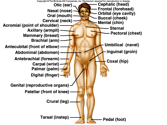
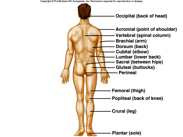
SURFACE ANATOMY
Knowledge of landmarks on the surface anatomy can aid in the identification of
internal structures. Below is listed the common name and corresponding
anatomical name. We will learn all of the anatomical names and more.
| Common name | Anatomical name | Common name | Anatomical name |
| head | Cephalic | fingers | Digital |
| skull | Cranial | navel | Umbilical |
| face | Facial | hip | Coxal |
| eye | Orbital | back | Dorsal |
| ear | Otic | loin | Lumbar |
| nose | Nasal | buttock | Gluteal |
| mouth | Oral | pubis | Pubic |
| neck | Cervical | thigh | Femoral |
| shoulder | Acromial | anterior knee | Patellar |
| chest | Thoracic | posterior knee | Popliteal |
| breast | Mammary | calf | Crural |
| armpit | Axial | ankle | Tarsal |
| upper arm | Brachial | toes | Digital |
| lower arm | Antebracial | sole of foot | Plantar |
| wrist | Carpal | heel | Calcaneal |
| palm | Palmar |
To see a picture, link to either the anterior or posterior views of the body.
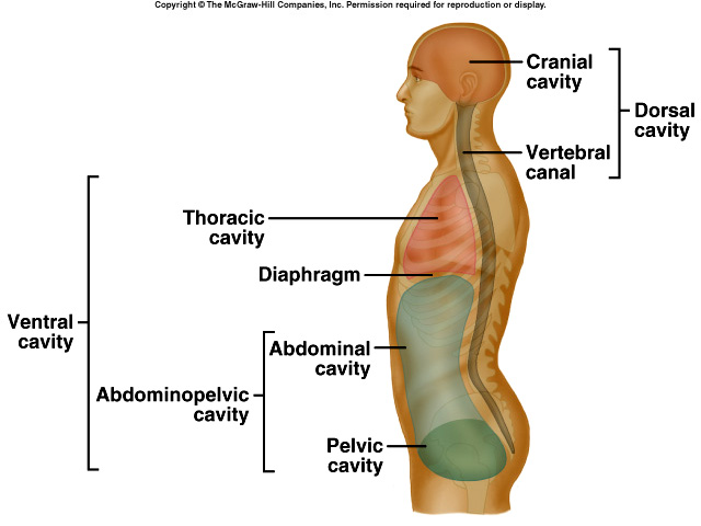
BODY CAVITIES AND MEMBRANES
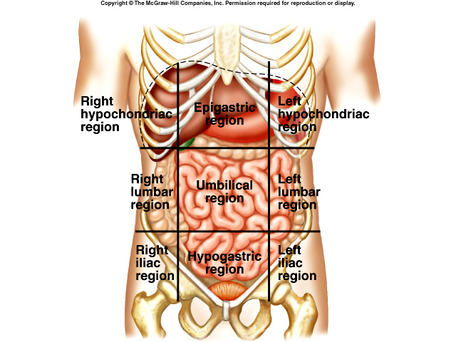 The body can be divided into a number of spaces called
cavities. These separate
the body into specific areas. The two main cavities are the dorsal cavity which
contains the cranial and vertebral cavities and the ventral cavity. The ventral
cavity contains the thoracic cavity (Right/Left pleural, pericardial) and the abdominopelvic cavity. The abdominopelvic cavity can be divided into nine regions
by drawing four lines. These regions are the right hypochondriac, epigastric, left
hypochondriac regions at the top; right lumbar, umbilical, left lumbar regions
in the middle; and right iliac, hypogastric (pubic), left iliac regions in the
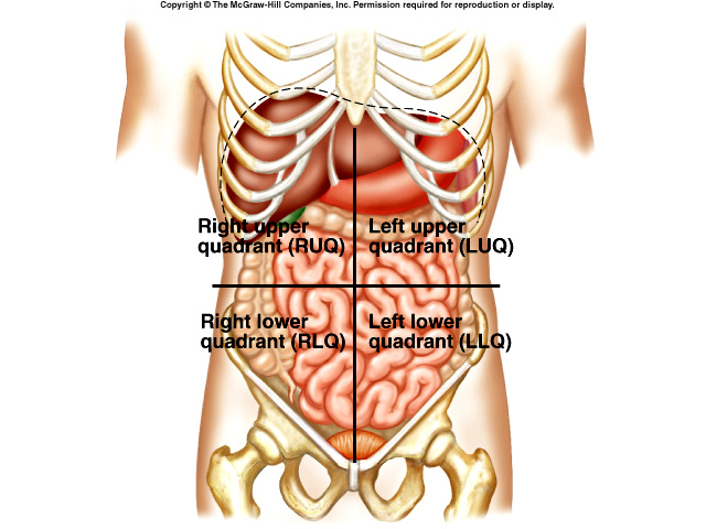
lower area. Sometimes the abdominopelvic region is divided into
four quadrants
by drawing a horizontal and vertical line through the umbilicus creating the
right and left upper lower quadrants.
The body can be divided into a number of spaces called
cavities. These separate
the body into specific areas. The two main cavities are the dorsal cavity which
contains the cranial and vertebral cavities and the ventral cavity. The ventral
cavity contains the thoracic cavity (Right/Left pleural, pericardial) and the abdominopelvic cavity. The abdominopelvic cavity can be divided into nine regions
by drawing four lines. These regions are the right hypochondriac, epigastric, left
hypochondriac regions at the top; right lumbar, umbilical, left lumbar regions
in the middle; and right iliac, hypogastric (pubic), left iliac regions in the
lower area. Sometimes the abdominopelvic region is divided into
four quadrants
by drawing a horizontal and vertical line through the umbilicus creating the
right and left upper lower quadrants.
Membranes
Mucous Membranes (mucosa) line body cavities that open directly to the exterior
of the body. They line the entire gastrointestinal tract, respiratory tract,
reproductive system, and most of the urinary tract. Basically they consist of an
epithelial layer that overlies the connective tissue. Serous membranes line
closed cavities and cover organs. Synovial membranes line the cavities of freely
moving joints.
ORGAN SYSTEMS
|
SYSTEM |
FUNCTION |
| Integumentary | Protection, water retention, thermoregulation, Vitamin D synthesis |
| Skeletal | Support, movement, protective enclosure of internal organs, blood formation, electrolyte and acid-base balance |
| Muscular | Movement, stability, control of body openings, heat production |
| Nervous | Internal electrical communication and coordination, sensation |
| Endocrine | Internal chemical communication and coordination |
| Cardiovascular | Distribution of nutrients, oxygen, wastes, hormones, electrolytes, heat, immune cells, and antibodies; fluid, electrolyte, and acid-base balance |
| Lymphatic | Recovery of excess tissue fluid, detection of pathogens, production of immune cells, defense |
| Digestive | Nutrient breakdown and absorption; liver has many functions |
| Respiratory | Absorption of oxygen, discharge of carbon dioxide, acid-base balance, speech |
| Excretory | Elimination of wastes, regulation of blood volume, control of electrolyte, fluid and acid-base balance |
| Reproductive | Continuation of the species |
THE CHEMICAL LEVEL
MATTER & ENERGY
Matter is defined as anything that occupies space and has mass.
Energy is defined as the capacity to do work. Energy can be differentiated as
potential (stored energy) or kinetic (energy in motion). Some kinds of energy
are chemical energy (released or absorbed in breaking or
forming atoms), radiant energy, and electrical energy.
All matter is made from elements. Known elements are listed on the Periodic
Table. An atom is the smallest particle of an element that has the properties of
that element whereas a molecule is a particle formed by the chemical union of
two or more atoms (O2). A compound is the combination of two or more
elements (H2O). Atoms join other atoms by forming bonds. When they
form these bonds, they gain, lose, or share electrons. Atoms that gain or lose
electrons become electrically charged and are called ions. Chemical reactions
form or break bonds and in doing so consume or generate energy.
Molecules containing carbon AND hydrogen atoms are organic and are usually non-electrolytes. The rest of the chemicals involved in the chemical reactions of the cell are inorganic. Of the INORGANIC COMPOUNDS that make up the body, the most important is Water. Other inorganic substances include carbon dioxide, oxygen, and some inorganic salts. Water is the most abundant substance in the body. It functions as a solvent and suspension media; it participates in chemical reactions; it releases heat slowly; it requires a large amount of heat to change from a liquid to a gas; and it serves as a lubricant. Oxygen, another inorganic molecule, is also necessary for life as it releases energy for metabolic activities.
ORGANIC compounds present in the cell
include carbohydrates (which serve as energy sources), lipids, and proteins (the
building blocks of the cell). Proteins are made from amino acids. The amino acid
sequence determines the proteins conformation which in turn determines its
function. Some proteins serve as enzymes. enzymes are catalysts in a living
system. They speed up chemical reactions without being consumed.
| ORGANIC MOLECULE | FUNCTION |
| CARBOHYDRATES | Mostly used as energy sources for cellular metabolism. Ex: glucose, glycogen |
| LIPIDS | Energy source, chemical messengers between cells, membrane component. |
| PROTEINS | Structure, communication between cells, membrane transport, catalysis, recognition and protection, movement, and cell adhesion |
What’s important about Acid-Base Balance?
THE CELLULAR LEVEL OF ORGANIZATION
The more hydrogen ions present in a solution the more acidic that solution and
the lower the pH. The body’s normal pH is 7.35 - 7.45 (slightly alkaline). To
maintain homeostasis, this pH must be maintained.
Maintenance is done through buffering systems; usually a weak acid or a weak
base that eliminates excess OH- or H+. We’ll discuss buffering systems later in
the chapter on acid – base balance.
Animal cells differ from plant cells in that animal cells do not have cell
walls. This allows much material to easily diffuse across the plasma membranes
into the cytosol of the cell where the materials can be stored as
inclusion bodies or can be utilized in the organelles of the cell.
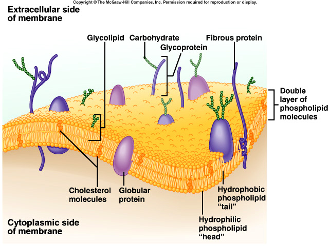
The PLASMA MEMBRANE is a very thin barrier that separates the internal
components of a cell from the external environment. The membrane is composed of
lipids (phospholipids, glycolipids, and cholesterol),
proteins (mostly glycoproteins) that form channels through which materials pass.
Proteins also act as transporters and serve as receptors, enzymes, cytoskeleton
anchor, and cell identity markers. The membrane
functions in cellular communication, as an electrochemical gradient which is
important for proper function of the cells. In selective permeability certain substances
are allowed to pass through while others are restricted.
Permeability depends on size of the molecule, lipid solubility, electric charge,
and the presence of transporters and channels.
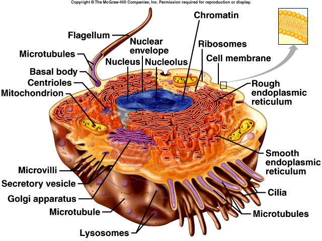
The Cell can be divided into two parts, the CYTOSOL or Intracellular Fluid
 and
the ORGANELLES. The cytosol is a semi-fluid portion of the cytoplasm in which
organelles and inclusions are suspended. The function
of the cytosol is to provide a place where metabolic reactions occur. The
organelles are specialized structures in the found in the cytosol. The most
important organelle is the Nucleus. It controls cellular activities,
contains the genetic material of the cell, which is seen as chromosomes during cellular
division, and is separated from the cytoplasm by a nuclear membrane. Within the
nucleus are nucleoli which are the site of assembly of ribosomes. Ribosomes have a role in protein synthesis. They are either free
floating in the cytosol or attached to the endoplasmic reticulum. The Endoplasmic Reticulum is
a network of membrane-enclosed channels continuous
with the nuclear membrane. Its function is to transport substances, store newly
synthesized molecules, detoxify chemicals, and release calcium ions involved in
muscle contractions. Another structure is the Golgi
Complex which processes, sorts, and delivers proteins and lipids to the plasma
membrane, lysosomes and secretory vesicles. Lysosomes contain digestive enzymes
that dissolve cellular contents and extracellular
materials. Mitochondria generate ATP (energy source of the cell). The
Cytoskeleton provides structure for the cell. Some cells contain Flagella and
Cilia which are external appendages use for locomotion. Finally, the Centrosome serves as mitotic spindles in dividing cells.
and
the ORGANELLES. The cytosol is a semi-fluid portion of the cytoplasm in which
organelles and inclusions are suspended. The function
of the cytosol is to provide a place where metabolic reactions occur. The
organelles are specialized structures in the found in the cytosol. The most
important organelle is the Nucleus. It controls cellular activities,
contains the genetic material of the cell, which is seen as chromosomes during cellular
division, and is separated from the cytoplasm by a nuclear membrane. Within the
nucleus are nucleoli which are the site of assembly of ribosomes. Ribosomes have a role in protein synthesis. They are either free
floating in the cytosol or attached to the endoplasmic reticulum. The Endoplasmic Reticulum is
a network of membrane-enclosed channels continuous
with the nuclear membrane. Its function is to transport substances, store newly
synthesized molecules, detoxify chemicals, and release calcium ions involved in
muscle contractions. Another structure is the Golgi
Complex which processes, sorts, and delivers proteins and lipids to the plasma
membrane, lysosomes and secretory vesicles. Lysosomes contain digestive enzymes
that dissolve cellular contents and extracellular
materials. Mitochondria generate ATP (energy source of the cell). The
Cytoskeleton provides structure for the cell. Some cells contain Flagella and
Cilia which are external appendages use for locomotion. Finally, the Centrosome serves as mitotic spindles in dividing cells.
| Organelle | Function |
| Nucleus | Surrounded by the nuclear membrane, this structure transmits and expresses genetic information. |
| Nucleolus | Found within the nucleus, this structure is the site of ribosome formation. |
| Ribosomes | Made of RNA and proteins, these structures make proteins. |
| Endoplasmic Reticulum | Rough ER has ribosomes attached and thus serves in the packaging of proteins that are to be secreted by the cell. Smooth ER lacks ribosomes and is the site of lipid synthesis. |
| Golgi Apparatus | Sorts and modifies the proteins that are to be secreted from the cell. |
| Mitochondria | Site that consumes Oxygen and produces carbon dioxide in the chemical process that transfers energy to ATP which can be used in cellular functions. |
| Cystoskeleton | Provides structure for the cell. |
| Cilia/Flagella | Used to facilitate movement of the cell or substances over the cell. |
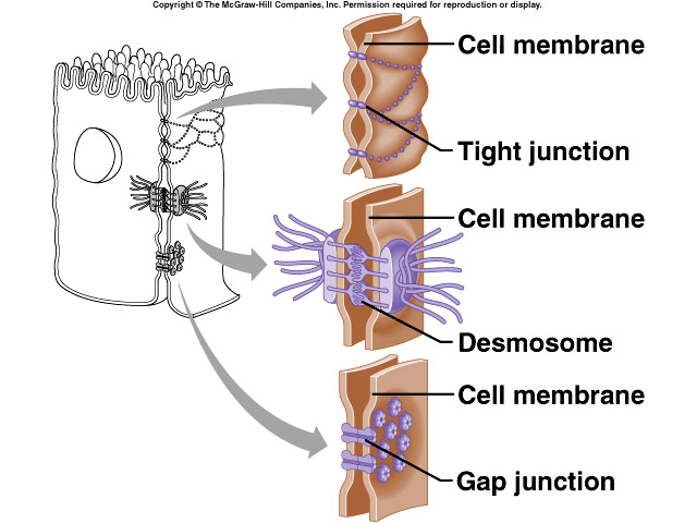
CELL JUNCTIONS are points of contact between adjacent plasma membranes.
One type is a tight
junction that forms fluid tight seals between cells. These are common in
epithelial cells that line the GI tract
and the urinary bladder. Another type is anchoring junctions that fasten cells
to one another or to extracellular material. They are found in tissues subjected to friction and
stretching (muscle tissue of the
heart). Lastly are communicating junctions which permit electrical or chemical
signals to pass from cell to
cell. These are found in heart muscle and smooth muscle of the GI tract.
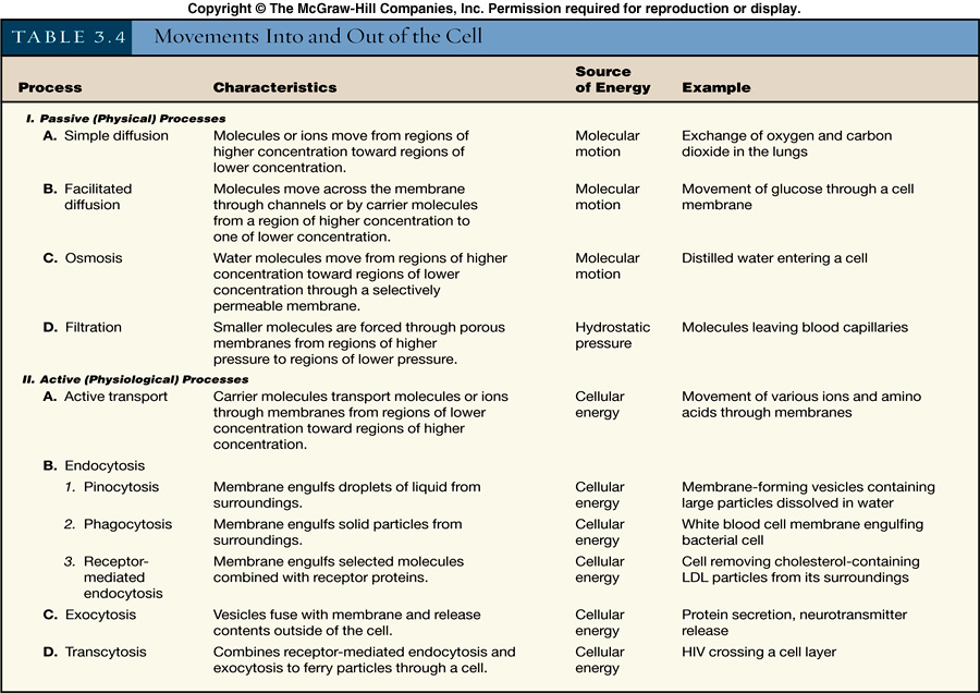
MOVEMENT OF MATERIALS ACROSS PLASMA MEMBRANES
The most important thing to remember about movement across membranes is that passive processes don’t use energy
whereas active processes do. Examples of passive processes include simple diffusion (particles
move from an area of high concentration to a lower concentration to reach a
point of equilibrium). This is seen in gas exchange in
tissues and lung. A second type is osmosis or the movement of a solvent through
a selectively permeable membrane. Here osmotic pressure, or the pressure needed
to stop the flow of water across the membrane,
depends on the permeability of the membrane and the tonicity of the solutions
involved (hypotonic, isotonic, or hypertonic). Osmosis always involves water. Filtration occurs when water and
some dissolved substances move across a membrane due
to gravity or hydrostatic water pressure. It always moves from higher levels of
pressure to lower levels. In facilitated diffusion certain molecules are helped
across a membrane by a transporter that moves the
molecules from a higher concentration gradient to a lower concentration.
Facilitated diffusion depends on difference in concentrations, number of
transporters available, and how quickly the transporter and the
substance combine.
Active Processes use energy from spitting ATP. Active transport can be either
primary active transport in which ATP directly moves a substance across the
membrane or secondary active transport in which energy
stored in ion differences drives the substance across the membrane. Another
active process is Bulk transport. Examples of this mechanism include endocytosis
(brining substances into a cell), phagocytosis (engulf a
substance and bringing that substance into the cell), pinocytosis (engulf tiny
droplet of extracellular fluid), receptor-mediated endocytosis (takes in
specific substances), and exocytosis (discharges substances from
cell).
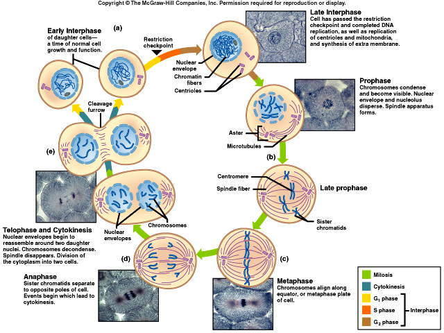
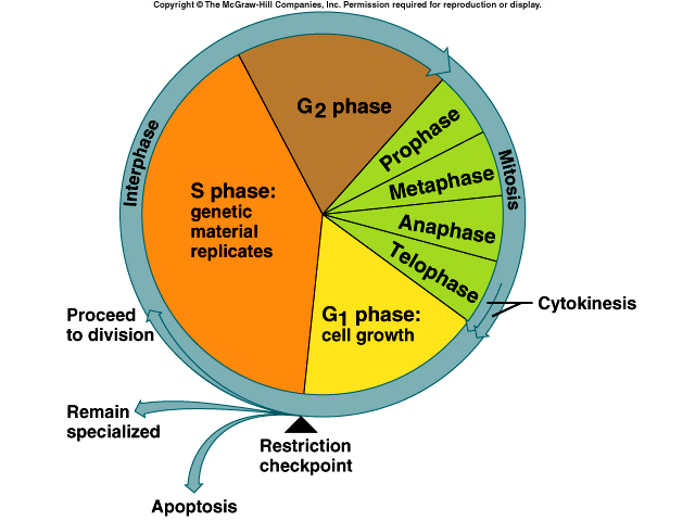
The process of NORMAL CELL DIVISION is a means by which cells replicate
themselves. It consists of a nuclear division (mitosis) and a cytoplasmic division (cytokinesis).
Somatic Cell Division results in an increase of body
cells. Parent cells and daughter cells both contain the diploid (2n) number of
chromosomes. Interphase is the time period between cell divisions during which
replication of DNA occurs and the original DNA molecule becomes two DNA
molecules. Some terms of mitotic cell division you should become familiar with
are:
-
prophase: chromatin material shortens and condenses
-
metaphase: centromeres of the chromatid spindles line up at the exact center of the mitotic spindle
-
anaphase: centromeres divide and chromosomes move to opposite ends of the cell
-
teleophase: nuclear envelope reappears and encloses chromosomes which resume chromatin form, nucleoli reappear, and mitotic spindle disappears
-
cytokinesis: division of the parent cell’s cytoplasm and organelles, cleavage appears to separate cytoplasm into usually two equal portions
The length of the cell cycle varies with location, temperature.
Meiosis or Reproductive Cell Division is a process that produces a haploid
number of chromosomes and consists of two nuclear divisions called reduction
division and equatorial division. In meiosis the homologous
chromosomes undergo synapsis (chromosomes become arranged in homologous pairs)
and crossing-over to result in two diploid
daughter cells. Then the two haploid daughter cells undergo mitosis (equatorial
division). The entire process results in four haploid daughter cells.
ABNORMAL CELL DIVISION: CANCER
Cancer is uncontrolled cell growth resulting in a tumor or neoplasm. A growth
than spreads (metastasis) is a malignant tumor. Non-spreading growth is called a
benign tumor. Cancer cells compete for nutrients and
space. They crowd out normal tissue until that normal tissue dies. Some causes
of cancer are carcinogens, which are found in the environment (such as chemicals
or radiation) and viruses. The treatment of cancer
employs a variety of methods depending on type of cancer. Some methods include surgical
removal, chemotherapy, radiation, immunotherapy to get rid of the
cancer. Bone marrow transplants may be used in certain
types of cancer to regenerate blood cells.
CELLULAR METABOLISM
Anabolism is the chemical reactions that combine simple substances into more
complex molecules (requires energy). For example, glycogenesis is the storage of
glucose as glycogen in the liver and skeletal muscles;
in glycogenolysis, glycogen is converted to glucose and released from the liver;
in gluconeogenesis protein or triglyceride molecules are converted into glucose;
and in lipogenesis glucose or amino acids are
converted into lipids.
Catabolism is the chemical reactions that breaks down complex molecules into
simpler ones (releases energy). During gycolysis, glucose breaks down into 2
molecules of pyruvic acid. In lipolysis, lipids break down into
glycerol and fatty acids.
Metabolic reactions require energy to start. Enzymes make cellular reactions
happen faster. Enzymes are usually proteins that promote specific reactions.
They are required in very small quantities and are not consumed in the reaction
process. Because they react on a specific substrate they are often referred to
as the "lock-and-key effect. Enzymes are usually named according to their
substrate with an -ase at the end.
Metabolic processes in cellular metabolism occur in three interconnected series of reactions: glycolysis, the citric acid cycle, and the electron transport chain. What is important about these reactions is that they produce the energy needed by in cellular respiration. The energy derived from this series yields thirty-eight ATPs.
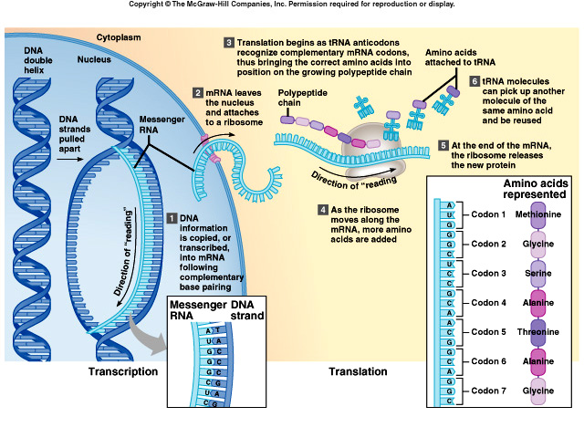
GENE ACTION: The function of cell is to make proteins and the instructions on how to make the proteins is found on the genes. The process is pretty simple. In transcription genetic information encoded in a region of the DNA helix is copied (transcribed) onto messenger RNA (mRNA). The next step is translation, a process by which the mRNA specifies the amino acid sequence of a protein in the ribosome. The result is an amino acid. Combinations of amino acids make different proteins. In summary, DNA → RNA → protein. (THIS IS GOSPEL – KNOW IT!)
Some more notes on NUCLEIC ACIDS AND PROTEIN SYNTHESIS
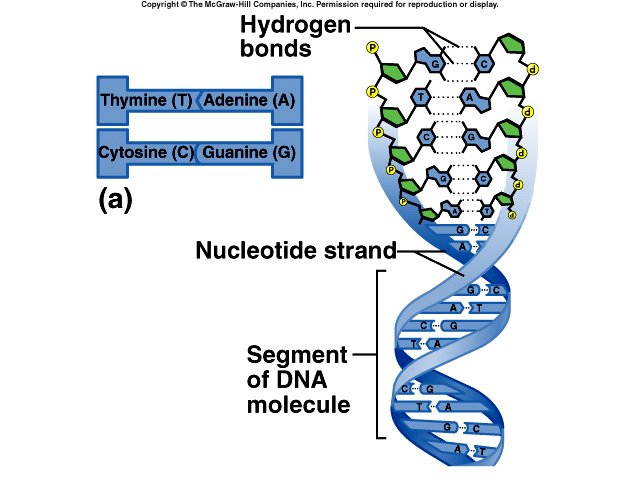
The total amount of DNA in a cell is called its genome. DNA is the blueprint
that tells the cell through RNA what proteins to make. DNA is composed of two
strands of nucleotides which contain Nitrogenous Bases
[purines (adenine & guanine) and pyrimidines (cytosine & thymine)].
Base pairing
rules dictate that they always, always pair up as A-T or C-G only. Also included
in DNA is a sugar, deoxyribose and a phosphate,
HPO4. The structure forms a double alpha helix (a twisted ladder formation).
Sections of DNA are the genes (a certain segment of DNA that contains the
necessary code to make a protein or RNA molecule). These
sections maintain the genetic code during reproduction, yet provide variability
due to crossing over during meiosis. DNA replication is the production of
identical strands of DNA. This must occur prior to cell
reproduction. Semiconservative replication means that each “old strand” of DNA
serves as a template upon which the “new strand” is synthesized. The double
strands separate to form two templates.
DNA relationship to proteins
The protein’s primary structure, that is the order and type of amino acids in
the chain determine its shape and function. The proteins that are produced
determine phenotype or the expression of a gene. And DNA is
the blueprint that tells the cell how to and which kinds of proteins to make.
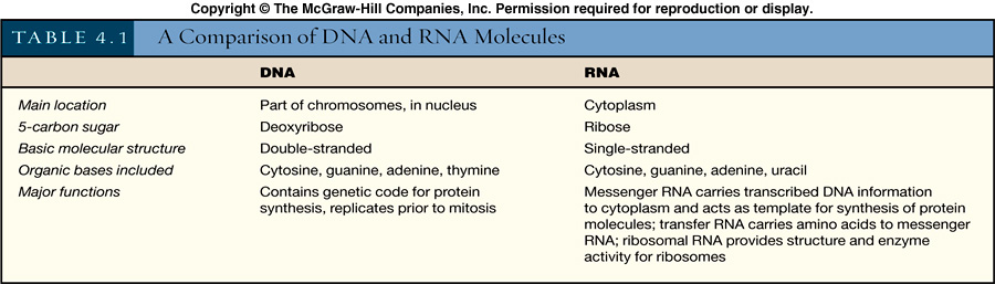
RNA Code
RNA is composed of single strands of nucleotides which contain Nitrogenous bases
[purines (adenine & guanine) and pyrimidines (cytosine and uracil)]. RNA also
contains a sugar, ribose, a HPO4. There are three
types of RNA, messenger RNA (mRNA) which is produced from DNA patterns in
transcription. The master DNA code is first copied onto mRNA through
transcription. Transfer RNA (tRNA) is also produced from DNA
patterns. Sixty-four varieties of codons are determined by anti-codons (nucleotide triplets)
and amino acid binding sites. 61/64 types represent some type of amino acid,
other types are either start or stop codons. Each variety of tRNA
converts the master code on mRNA into a specific amino acid. Ribosomal RNA (rRNA)
forms the major part of the ribosome and participates in protein synthesis. Link
for a comparison of DNA and RNA molecules.
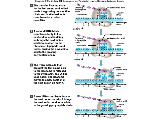
Review of Steps in Protein Synthesis Sequence
-
DNA unzips
-
Transcription produces mRNA using the DNA code by complimentary base pairing mRNA attaches to ribosome
-
tRNA anticodons attach to complimentary codons of mRNA amino acids join to produce protein
-
Translation: production of a protein from a mRNA strand
a. All elements needed to synthesize a protein are brought together on the ribosome.
Mutation Mechanisms or Nucleic acids gone bad
A Mutation is a permanent change in the DNA that may be passed along from
generation to generation. The wild type (strain) of the organism exhibits,
non-mutated characteristics. A mutant strain can show variance
in morphology, nutritional characteristics, genetic control mechanisms,
resistance to chemicals, temperature preference, and any type of enzymatic
function.
Causes of mutation can be spontaneous where there is a random change in DNA.
This arises from mistakes in DNA replication. Mutations can be induced due to chemical
or physical factors. Many chemical mutations are also
carcinogenic and can result in cell death. The categories of mutations are as
follows:
-
Point mutations change the nature of one gene. These can be either frameshift (deletion or insertion of a base pair) or substitution in which the wrong base pair is put in place of correct bp producing error in base pairing, thus a change in the codon.
-
Inversion is the change in one or two codons (adjacent base pairs change position). These can be silent (no change in amino acid) if the same bp are exchanged or missense which can have consequences of none to severe. This is due to a faulty, nonfunctional protein, a different, but functional protein, or there can be no significant alteration in protein function.
-
Some mutation result in nonsense or STOP codon. The protein stops being produced. If large mutations in which whole chromosomes are lost or large genetic sequences are inserted, the cell or organism often will not survive. Thus major mutations alter the number of genes present.
TISSUE LEVEL OF ORGANIZATION
TYPES OF TISSUES AND THEIR ORIGINS
A tissue is a group of similar cells that usually have the same embryological
origin and are specialized for a particular function. In a tissue surrounding
the cells is fluid called extracellular fluid. There are two types of
extracellular fluid: interstitial fluid and plasma. Plasma is found in the blood
stream and serves as a transport medium for the blood cells. In addition plasma
carries nutrients, proteins, electrolytes, gases and wastes.
-
Epithelial tissue is a specific type of tissue that covers body surfaces, lines hollow organs, body cavities, and ducts; forms glands.
-
Connective tissue: protects and supports the body and its organs; binds organs together; stores energy reserves as fat; provides immunity.
-
Muscle tissue: excitable cells responsible for movement and generation of force.
-
Nervous tissue: excitable cells that initiate and transmit action potentials that help coordinate body activities.
EPITHELIAL TISSUE
Epithelial tissue is composed mostly of cells with little extracellular material and these
cells are closely packed together
and are arranged in continuous sheets in one or multiple layers. The apical
surface is exposed to body cavity, lining of an internal organ or
the exterior of the body and a basal surface which is attached to the basement
membrane (two layers: basal and reticular lamina). Numerous cell junctions are
present to secure cells to each other. Epithelial tissue is avascular (not supplied by blood vessels). It gets nutrients and removes wastes
by diffusion. Epithelial tissue can be renewed easily and has a nerve supply.
The functions of epithelial tissue include protection, filtration, lubrication,
secretion, digestion, absorption, transportation, excretion, sensory reception,
and reproduction. Arrangement of cells can be simple (one layer), stratified
(cells stacked), or pseudostratified . The shapes can be
squamous (flattened and scalelike), cuboidal (cube shaped), columnar (tall and
cylindrical), or transitional.
Each type found has a different function. For example, simple squamous
epithelium functions in filtration and diffusion. Simple cuboidal epithelium and
non-ciliated simple columnar epithelium function in secretion and
absorption. Ciliated columnar epithelium move fluids or particles along
passageways by ciliary action. Stratified squamous and cuboidal epitheliums
provide protection. Whereas stratified columnar epithelium provides
both protection and secretion. Transitional epithelium permits distention.
Pseudostratified columnar epithelium allows secretion and movement of mucous by
ciliary action. The most widespread epithelium in the body is stratified
squamous epithelium.
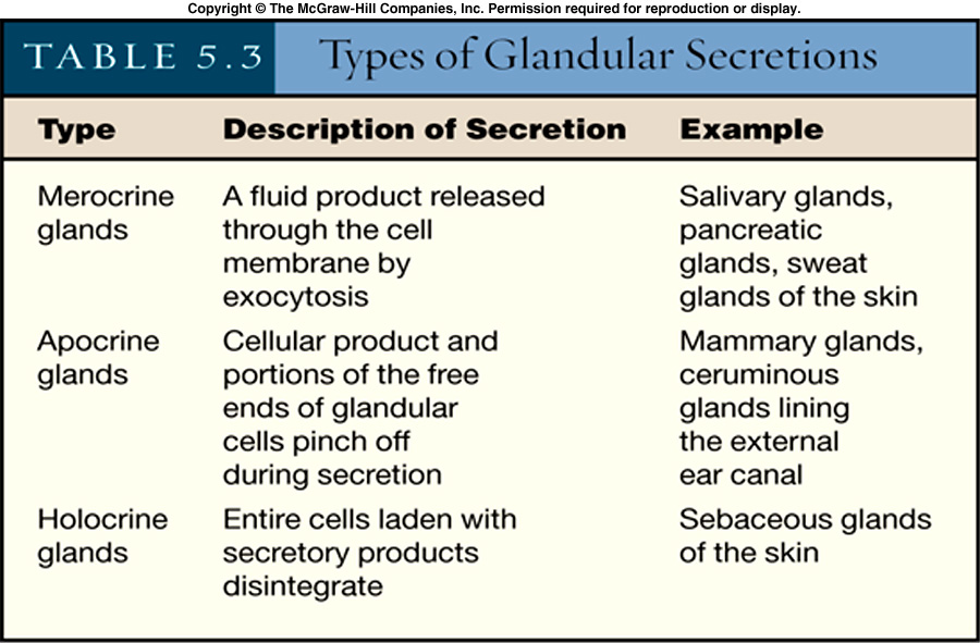
Glandular Epithelium
Exocrine glands secrete their products into ducts and are either unicellular or multicellular. Functional types
include holocrine glands (oil gland of the skin), merocrine gland which
discharge product by exocytosis,
and apocrine glands (mammary glands).
The endocrine glands are ductless, secrete products into extracellular fluid and
into blood.
CONNECTIVE TISSUE
Connective tissue is the most abundant body tissue in the body. It consists of
widely separated cells, ground substance and fibers which form a matrix. It does not occur on
free surfaces. Except for cartilage and tendons, it has a nerve
supply and usually has a good blood supply.
Types of connective tissue cells found in the body include fibroblasts which
secrete the molecules that form the matrix, macrophages which are derived from a
monocyte and used to phagocytize other dead cells,
plasma cells which make and secrete antibodies, mast cells which produce
histamine, adipocytes (fat cells) and leukocytes (white blood cells). Connective
tissue serves to connect different tissues together. Other functions include
physical protection, immune protection, movement, heat production, and
transport.
The connective tissue matrix is comprised of ground substance and fibers. The
fibers in the matrix provide the support and strength for tissues. Types of
fibers include collagen fibers, elastic fibers, reticular fibers.
Types of Mature Connective Tissue:
-
loose connective tissue: fibers loosely woven
-
dense connective tissue: contains more fibers and less cells than loose connective tissue
-
areolar connective tissue: widely distributed in the body, combined with adipose tissue, it forms the subcutaneous layer
-
reticular connective tissue: helps to bind together the cells of smooth muscle
-
adipose tissue, composed of adipocytes which store fats
-
dense regular connective tissue: provides strong attachment between various structures (tendons and ligaments)
-
dense irregular connective tissue: provides strength (fasciae)
-
elastic connective tissue: allows stretching off various organs (lungs)
Cartilage
Hyaline cartilage is the most abundant type of cartilage in the body. In muscle
tissue it is the gristle. It affords flexibility and support, reduces friction
and absorbs shock. Fibrocartilage provides support and fusion.
Elastic cartilage gives support, yet maintains shape (epiglottis).
Bone tissue (osseous), blood (vascular tissue), muscular, and nervous tissue will be
covered at a later time.
INTEGUMENTARY SYSTEM
SKIN
 Skin is an organ because it consists of different tissues that are joined to
perform specific activities. It covers about 2 sq. meters and weighs 10-11 lb.
The range in thickness is from 0.5 to 4.0 mm depending on its
location.
Skin is an organ because it consists of different tissues that are joined to
perform specific activities. It covers about 2 sq. meters and weighs 10-11 lb.
The range in thickness is from 0.5 to 4.0 mm depending on its
location.
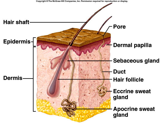
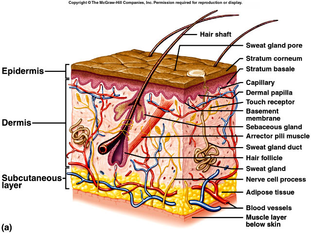
The anatomy of skin includes three layers; the epidermis is the most outer
layer, a thinner portion that is composed of epithelium. Next is the dermis, the
inner, thicker portion that is composed of connective tissue.
Beneath the dermis is a subcutaneous layer which is composed of superficial
fascia (hypodermis).
|
Functions of Skin |
| Regulation of body temperature; evaporation of sweat lowers elevated body temperature, however most heat is lost by radiation. |
| Protection from abrasions, disease, dehydration |
| Sensation: touch, pressure, temperature, and pain. |
| Excretion: removal of wastes (Carbon dioxide) |
| Immunity |
| Blood reservoir |
| Synthesis of vitamin D |
| Social functions |
The epidermis is composed of stratified squamous epithelium. Cell types found in the epidermis and their function are: the keratinocytes that produce keratin which waterproofs the skin, the melanocytes that produce the pigment melanin which absorbs UV light, the Langerhans cell that arise in the bone marrow and migrate to the epidermis and help with immunity, and Merkel cells that are thought to function in the sensation of touch.
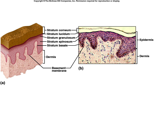
The layers of the epidermis from deepest to most superficial are:
-
stratum basale: single layer of cuboidal to columnar cells which are stem cells (that produce the keratinocytes), melanocytes, and tactile cells.
-
stratum spinosum: layer contains 8 -10 rows of many-sided keratinocytes
-
stratum granulosum: 2-5 layers of keratinocytes that develop keratohyalin (precursor of keratin) which functions in waterproofing the skin
-
stratum lucium: normally seen only in thick skin
-
stratum corneum: uppermost 25-30 layers of cells completely filled with keratin; keratinization occurs in cellular development as the cells are pushed towards the surface.
The dermis is composed of connective tissue containing collagen and elastic
fibers. The papillary region, the outer 1/5, has a surface area that is
increased by projections called dermal papillae (ridges, that when
formed cause the epidermal layer to develop finger prints) which are filled with
capillaries and tactile receptors. The reticular region, or inner portion
consists of dense irregular connective tissue containing collagen,
adipose tissue, hair follicles, sebaceous (oil) glands, nerves and sudoriferous
(sweat) glands. Beneath the dermis is the subcutaneous layer (hypodermis). This
layer binds the skin to underlying tissue, pads the body, and serves as an
energy reserve.
Skin color is due to melanin, carotene (yellow), and hemoglobin (red). All races have about the
same number of melanocytes, it’s the amount of melanin produced that determines
the skin color. UV light stimulates melanin production.
EPIDERMAL DERIVATIVES (ACCESSORY ORGANS OF THE SKIN)
Hair
The primary function of hair is protection. It decreases heat loss and protects
us from large foreign particles being inhaled and getting into our eyes and ears. The anatomy consists of a shaft above
the surface, a root that penetrates the dermis and
subcutaneous layer, and a hair follicle. Sebaceous (oil) glands, arrector pili
muscles, hair root plexus are also associated with hair. Hair color is primarily due
to melanin.
Nails
Nails are hard, keritinized epidermal cells over the dorsal surface of the
terminal portions of fingers and toes. Parts of a nail are the body free edge,
root, lunula, eponychium (cuticle) and the nail matrix. The function
is protection and to help us grasp small objects.
Glands
Sebaceous (oil) glands are usually connected to hair follicles, produce sebrum
which moistens hair and waterproofs skin. An infection of sebaceous glands leads
to boils or pimples.
Sudoriferous (sweat) glands are basically of two types: apocrine which are found
in the skin of the axilla, the pubic region and areolae and are stimulated
during emotional stress or sexual excitement and merocrine (eccrine)
sweat glands which cover most of the body. The ducts terminate at a pore on the
surface of the epidermis. The function is to produce perspiration and remove
small amounts of waste. Ceruminous glands are modified
sudoriferous glands that secrete cerumen (ear wax) which helps prevent foreign
particles from entering the ear. Mammary glands produce milk in lactating
females after childbirth.
SKIN AND HOMEOSTASIS
The major function of the skin is the maintenance of normal body temperature
(thermoregulation). Heat is lost or given off by radiation, conduction,
convection, and evaporation. If body temperature is high, stimuli are sent to the brain
which responds by sending stimuli back to the skin to
produce sweat and for the blood vessels to vasodilate. As sweat evaporates,
large amounts of heat energy leave the body.
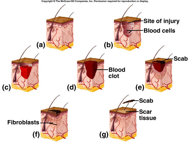
Skin Wound Healing
Epidermal wounds are repaired by enlargement and migration of basal cells,
contact inhibition, and division of migrating and stationary basal cells. In
deep wound healing there is an inflammatory phase where blood
clot unites the wound edges, epithelial cells migrate across the wound, and
vasodilatation delivers phagocytes. In the migratory phase, epithelial cells
beneath the scab bridge the wound, fibroblasts develop scar
tissue and damaged blood vessels repair themselves. The proliferative phase
continues more of the above. In the maturation phase, the scab sloughs off,
blood vessels are repaired. Finally in fibrosis there is the
process of scar tissue formation.
DISORDERS
Skin Cancer is usually a result of excessive sun exposure. Basal cell carcinoma
is the most common and rarely metastasizes. Squamous cell carcinoma has a
variable tendency to metastasize and develops from
preexisting lesions on sun exposed skins. Malignant melanomas arise from
melanocytes, readily metastasize, and can cause death within months especially
if not treated. It is mostly seen in young women.
Predisposing factors include skin type (fair skin more susceptible), amount of
skin exposure, family history, age and immunologic status.